
human anatomy and physiology laboratory manual answer key pdf
This laboratory manual provides a comprehensive guide for exploring human anatomy and physiology through hands-on activities and detailed exercises. It includes visual aids, practical experiments, and key concepts to enhance learning. The accompanying answer key offers solutions to exercises, enabling students to assess their understanding and improve retention of complex topics effectively.
Overview of the Laboratory Manual
This laboratory manual is designed to complement anatomy and physiology coursework, offering structured exercises and activities to reinforce theoretical concepts. It includes detailed visuals, dissection guides, and practical experiments to enhance hands-on learning. The manual is divided into sections corresponding to major body systems, ensuring a logical progression of topics. Each chapter features clear objectives, materials lists, and step-by-step instructions to facilitate understanding. Accompanying answer keys provide correct responses to exercises, enabling students to self-assess and improve their grasp of complex anatomical and physiological principles. The manual also integrates digital resources, such as virtual simulations and review questions, to support diverse learning styles and promote academic success.
Importance of Lab Manuals in Anatomy and Physiology Education
Lab manuals are essential for anatomy and physiology education, providing structured, hands-on experiences that enhance understanding of complex biological concepts. They bridge the gap between theoretical knowledge and practical application, allowing students to explore human structures and functions through dissections, experiments, and observations. These manuals foster active learning, engagement, and critical thinking, while also improving retention of key concepts. Additionally, lab manuals serve as valuable resources for self-study and review, often including answer keys to guide students and reinforce their learning outcomes. This practical approach ensures a deeper appreciation and mastery of human anatomy and physiology.
The Skeletal System
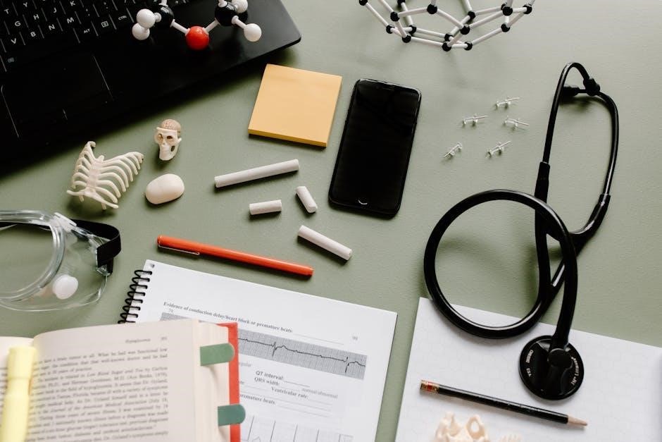
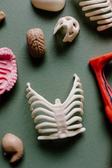
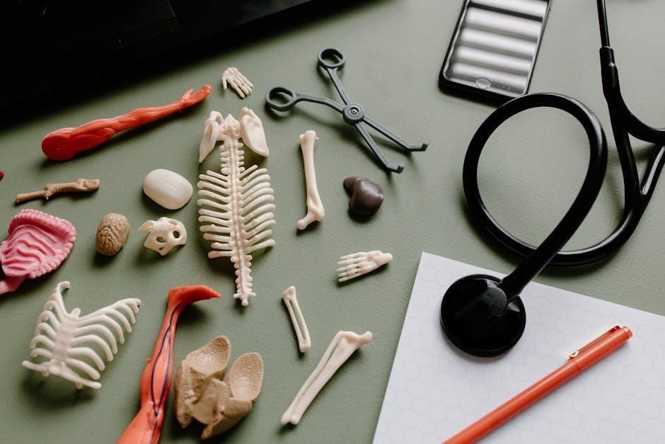
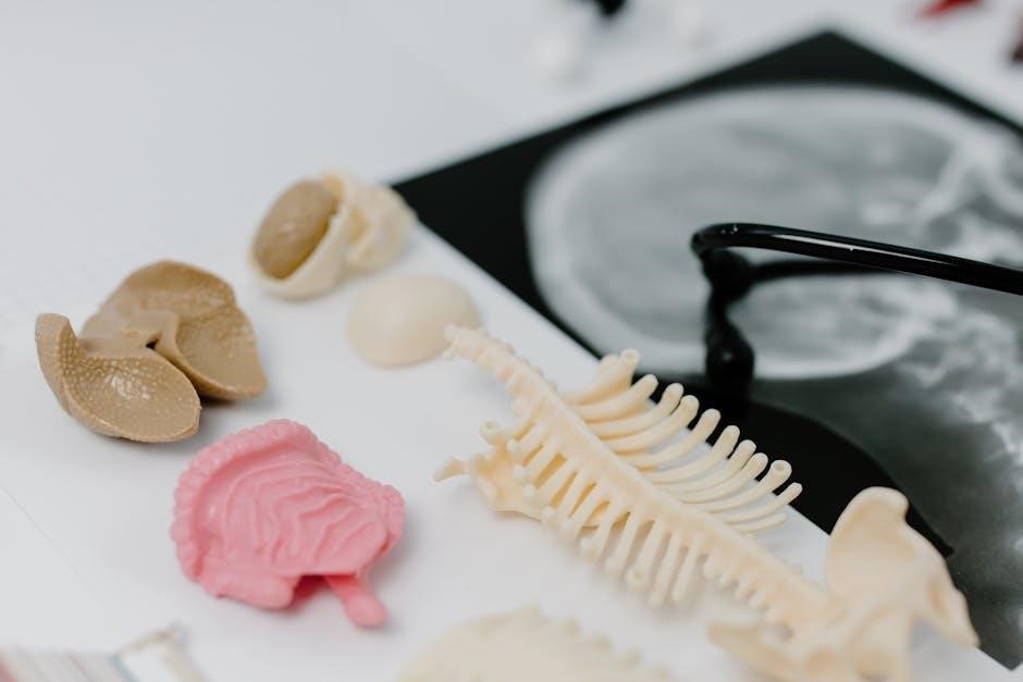
The skeletal system comprises bones, joints, and ligaments, providing structural support, protection, and movement. It is studied through lab manuals for detailed anatomical understanding.
Structure and Function of Bones
Bones are dynamic tissues composed of organic and inorganic materials, primarily collagen and calcium phosphate, providing strength and flexibility. The periosteum, a fibrous membrane, covers bones, while bone marrow produces blood cells. Bones function as structural support, enabling movement and protecting vital organs like the brain and heart. They also store minerals such as calcium and phosphorus for bodily functions. Different bone types—long, short, flat, irregular, and sesamoid—perform specialized roles, like weight-bearing and protection. This section explores bone anatomy, growth, and remodeling processes, essential for understanding human anatomy and physiology.
Classification of Joints and Their Movements
Joints are classified based on their ability to move and the type of connective tissue they contain. Fibrous joints are immovable, found between bones like the skull sutures. Cartilaginous joints allow limited movement, such as the intervertebral discs. Synovial joints are highly movable, like the shoulder and knee, and contain synovial fluid for lubrication. Movements include flexion, extension, abduction, adduction, rotation, and circumduction. Understanding joint classification and their range of motion is crucial for analyzing human movement and skeletal function. This section details the structure and types of joints, enhancing comprehension of musculoskeletal mechanics.
Axial vs. Appendicular Skeleton
The axial skeleton forms the body’s central framework, comprising the skull, vertebral column, ribs, and sternum. It provides structural support, protects vital organs like the brain and heart, and serves as an attachment point for muscles. In contrast, the appendicular skeleton includes the upper and lower limbs, shoulders, hips, and pelvic girdles. Its primary functions are movement, muscle attachment, and facilitating locomotion. Together, these systems enable a wide range of motion and stability, forming the foundation of human anatomy. Understanding their differences is essential for analyzing posture, movement, and overall skeletal functionality in various physiological contexts.
The Muscular System
The muscular system consists of skeletal, smooth, and cardiac muscles, enabling movement, maintaining posture, and facilitating bodily functions like digestion and circulation. It works synergistically with the skeletal system to produce motion and maintain stability, while also playing a crucial role in overall physiology.
Types of Muscles: Skeletal, Smooth, and Cardiac
The muscular system comprises three distinct types of muscles: skeletal, smooth, and cardiac. Skeletal muscles are voluntary, attached to bones, and responsible for movement and posture. Smooth muscles are involuntary, found in internal organs, and facilitate functions like digestion. Cardiac muscle is specialized for the heart, enabling continuous, rhythmic contractions. Each type varies in structure, function, and control mechanisms, working together to maintain bodily functions and overall mobility. Understanding their differences is crucial for comprehending human physiology and addressing related disorders. This section provides detailed insights into their characteristics and roles in the body.
Muscle Physiology: Contraction and Relaxation
Muscle contraction and relaxation are fundamental physiological processes governed by the sliding filament theory. Contraction occurs when actin and myosin filaments interact, driven by ATP hydrolysis and calcium ion binding to troponin. This process enables movement, maintains posture, and supports bodily functions. Relaxation follows when calcium ions are pumped back into the sarcoplasmic reticulum, allowing tropomyosin to block myosin binding sites. These mechanisms are essential for voluntary and involuntary muscle activities, ensuring precise control over movement and tension. Understanding these processes is vital for appreciating muscle function and addressing related disorders. This section explores the intricate mechanisms behind muscle physiology in detail.
Major Muscle Groups and Their Functions
The human body contains several major muscle groups, each with distinct functions. The pectoralis major facilitates shoulder flexion and extension, while the latissimus dorsi enables shoulder adduction and extension. Deltoids are crucial for shoulder abduction and flexion. The biceps brachii and triceps brachii work together to flex and extend the elbow. Quadriceps and hamstrings control knee movements, with quadriceps extending and hamstrings flexing the knee. Gluteal muscles, including the gluteus maximus, are essential for hip extension and external rotation. These muscle groups collectively enable movement, maintain posture, and support daily activities, making them vital for overall bodily function and mobility. Understanding their roles is key to appreciating human physiology.
The Circulatory System
The circulatory system, comprising the heart, blood, and blood vessels, transports oxygen, nutrients, and hormones throughout the body. It maintains homeostasis and supports immune function.
Components of Blood and Their Functions
Blood is a connective tissue consisting of plasma, red blood cells (RBCs), white blood cells (WBCs), and platelets. Plasma, the liquid portion, transports nutrients, hormones, and waste products. RBCs, containing hemoglobin, carry oxygen throughout the body. WBCs are vital for immune defense, fighting infections and diseases. Platelets play a crucial role in blood clotting, preventing excessive bleeding from injuries. Together, these components ensure proper bodily functions, maintaining homeostasis and overall health. Understanding their roles is essential for diagnosing and treating various medical conditions.
Structure and Function of the Heart
The heart is a muscular organ divided into three layers: the epicardium (outer layer), myocardium (muscular layer), and endocardium (inner layer). It has four chambers: the right and left atria, and the right and left ventricles. Blood flows through the heart in a single direction due to valves. The right side receives deoxygenated blood, which is pumped to the lungs for oxygenation. The left side receives oxygenated blood and distributes it to the body. The heart’s contractions are regulated by the sinoatrial node and the Bundle of His, ensuring efficient circulation of blood throughout the body.
Arteries, Veins, and Capillaries: Key Differences
Arteries carry oxygenated blood away from the heart, while veins return deoxygenated blood to it. Arteries have thick, elastic walls to withstand high blood pressure, whereas veins have thinner walls and one-way valves to prevent backflow. Capillaries are microscopic vessels where oxygen, nutrients, and waste products are exchanged between blood and tissues. Arteries and veins are structurally different, with arteries having more smooth muscle and elastic fibers. Capillaries, the smallest vessels, allow for diffusion of substances due to their thin walls. Understanding these differences is crucial for studying circulation and blood flow dynamics in the human body.

The Nervous System
The nervous system controls body functions, enabling communication through electrical and chemical signals. It consists of the central nervous system (CNS) and peripheral nervous system (PNS), coordinating sensory input, motor responses, and internal regulation to maintain homeostasis and overall bodily functions effectively.
Structure and Function of Neurons
Neurons, or nerve cells, are specialized cells designed to transmit information through electrical and chemical signals. A neuron consists of three main parts: dendrites, which receive signals; the cell body, containing the nucleus and organelles; and the axon, a long extension that transmits signals to other neurons, muscles, or glands. The synapse is the gap between neurons where chemical neurotransmitters are released, allowing communication. Neurons play a crucial role in controlling voluntary and involuntary body functions, enabling sensory perception, movement, and maintenance of homeostasis. Understanding their structure and function is essential for studying the nervous system’s intricate operations and regulatory processes.
Types of Nervous Tissue and Their Roles
Nervous tissue consists of two main types: gray matter and white matter. Gray matter contains neuron cell bodies and is primarily found in the brain, spinal cord, and ganglia, playing a key role in information processing. White matter, composed of myelinated axons, facilitates rapid signal transmission between neurons. Nervous tissue supports the nervous system’s functions, including controlling voluntary and involuntary actions, regulating body functions, and enabling thought and communication. It allows for efficient transmission of electrical and chemical signals, crucial for the body’s ability to respond to stimuli and maintain homeostasis.
Reflexes and Nerve Impulses
A reflex is an involuntary, automatic response to a stimulus, involving a neural pathway called a reflex arc. It consists of a sensory neuron detecting the stimulus, a motor neuron initiating a response, and sometimes an interneuron for processing. Nerve impulses, or action potentials, are electrical and chemical signals that transmit information along neurons. They occur due to ion concentration changes across cell membranes, enabling rapid communication within the nervous system. Reflexes ensure quick reactions, such as withdrawing a hand from heat, while nerve impulses are essential for controlling body functions, movement, and thought processes.
The Digestive System
The digestive system includes organs like the mouth, esophagus, stomach, intestines, and liver, responsible for breaking down food into nutrients through processes like ingestion, digestion, and absorption, excreting waste.
Structure and Function of the Digestive Tract
The digestive tract, also known as the alimentary canal, is a continuous passageway from the mouth to the anus. It includes the mouth, esophagus, stomach, small intestine, and large intestine. Each section specializes in specific digestive processes. The mouth initiates mechanical digestion with teeth and enzymes, while the esophagus transports food to the stomach via peristalsis. The stomach secretes digestive acids and enzymes to break down food into a liquid mixture called chyme. The small intestine absorbs nutrients into the bloodstream through finger-like projections called villi. Finally, the large intestine absorbs water and forms waste for elimination. This system ensures efficient nutrient extraction and waste removal.
Role of Digestive Enzymes
Digestive enzymes are biological catalysts that break down complex food molecules into smaller, absorbable nutrients. They are produced in glands like the pancreas and salivary glands and work in specific sections of the digestive tract. For instance, amylase in the mouth and small intestine breaks down carbohydrates into glucose, while proteases like pepsin and trypsin digest proteins into amino acids. Lipases, such as pancreatic lipase, break down fats into fatty acids and glycerol. Enzymes ensure efficient nutrient absorption, and their deficiency can lead to malnutrition or conditions like lactose intolerance. Each enzyme targets specific substrates, optimizing the digestion process for energy and cellular function.
Processes of Ingestion, Digestion, and Absorption
Ingestion begins with consuming food, which is mechanically broken down by chewing. Digestion involves both mechanical and chemical processes, breaking food into smaller molecules. Enzymes in saliva, stomach acid, and pancreatic juice chemically degrade carbohydrates, proteins, and fats. Absorption occurs primarily in the small intestine, where specialized villi increase surface area, allowing nutrients to enter the bloodstream. Nutrients are then transported to cells for energy, growth, and repair. This sequential process ensures efficient nutrient extraction, maintaining bodily functions and overall health. Each step is essential for converting ingested food into usable energy and sustain life.
The Respiratory System
The respiratory system enables gas exchange through inhalation and exhalation. Air enters via the nose, trachea, and bronchi, reaching alveoli for oxygen absorption.
Structure of the Lungs and Airway
The respiratory pathway begins with the nose or mouth, leading to the pharynx, larynx, and trachea. The trachea divides into bronchi, entering the lungs. Each lung contains lobes, with the right lung having three and the left having two. Bronchi branch into smaller bronchioles, ending in alveoli, where gas exchange occurs. The lungs are protected by the ribcage and diaphragm, facilitating expansion during breathing. This intricate structure ensures efficient oxygen intake and carbon dioxide removal, maintaining proper respiratory function and overall health.
Mechanism of Breathing: Inhalation and Exhalation
Breathing involves the coordinated action of the diaphragm and intercostal muscles. Inhalation occurs as the diaphragm contracts and flattens, increasing chest cavity volume and lowering intrathoracic pressure. Air rushes in through the airways into alveoli for gas exchange. Exhalation is passive, driven by elastic recoil of lungs and chest wall. During forced exhalation, abdominal and internal intercostal muscles assist. This mechanism ensures continuous oxygen supply and carbon dioxide removal, vital for cellular respiration and maintaining pH balance in blood. Proper breathing is essential for overall physiological function and health.
Gas Exchange and Oxygen Transport
Gas exchange occurs in alveoli, where oxygen diffuses into blood and carbon dioxide diffuses out. Oxygen binds to hemoglobin in red blood cells, forming oxyhemoglobin, and is transported to tissues. Myoglobin in muscle cells stores oxygen for later use. This process is vital for cellular respiration, enabling energy production. Efficient oxygen transport ensures proper organ function and maintains acid-base balance. Understanding these mechanisms is crucial for diagnosing respiratory and circulatory disorders, emphasizing the importance of studying anatomy and physiology in laboratory settings to grasp these complex processes effectively.
The Urinary System
The urinary system consists of kidneys, ureters, bladder, and urethra, functioning to filter waste, regulate electrolytes, and produce urine. It maintains homeostasis and overall health;
Structure and Function of the Kidneys
The kidneys are bean-shaped organs located in the lower back, responsible for filtering blood to remove waste and excess substances. They regulate electrolyte balance, blood pressure, and pH levels. Each kidney contains a renal cortex and medulla, with nephrons as the functional units. Nephrons filter blood, reabsorb nutrients, and produce urine. The kidneys also produce hormones like erythropoietin and renin, essential for red blood cell production and blood pressure regulation. Proper kidney function is vital for maintaining homeostasis, with diseases like diabetes and hypertension potentially causing damage. Regular lab tests, such as urinalysis, help assess kidney health and function.
Process of Urine Formation
The process of urine formation begins with filtration in the glomerulus, where blood is filtered to produce filtrate. This filtrate then moves through the renal tubules, where essential nutrients, water, and ions are reabsorbed back into the bloodstream. Tubular secretion removes additional waste and excess substances from the blood into the filtrate. The remaining filtrate, now urine, is concentrated and collected in the renal pelvis before being transported to the bladder via the ureters. This process ensures the elimination of waste while maintaining proper electrolyte and fluid balance in the body.
Role of the Ureters, Bladder, and Urethra
The ureters are narrow, muscular tubes that transport urine from the kidneys to the bladder for storage. The bladder, a hollow, elastic organ, stores urine until it is expelled from the body. The urethra, a tube leading from the bladder to the exterior, serves as the passageway for urine during urination. The urethral sphincter regulates the release of urine, ensuring proper control. Together, these structures form the excretory pathway of the urinary system, enabling waste removal while maintaining continence and overall urinary health.
The Reproductive System
The reproductive system is essential for producing offspring, involving male and female organs that support gamete production, fertilization, and development of embryos into viable organisms.
Male and Female Reproductive Organs
The male reproductive system includes the testes, penis, and accessory glands like the prostate and seminal vesicles, which produce and deliver sperm. The female reproductive system consists of the ovaries, uterus, vagina, and mammary glands. The ovaries produce eggs, while the uterus supports embryonic development. The vagina serves as the birth canal and pathway for menstrual flow. Both systems are designed to facilitate reproduction, with males producing sperm and females providing the environment for fertilization and fetal growth. Understanding these organs is crucial for comprehending reproductive health and processes.
Functions of Hormones in Reproduction
Hormones play a vital role in regulating reproductive processes. In males, testosterone promotes sperm production and secondary sexual characteristics. In females, estrogen and progesterone control the menstrual cycle, pregnancy, and breast development. The pituitary gland releases gonadotropins, such as follicle-stimulating hormone (FSH) and luteinizing hormone (LH), which stimulate the gonads to produce hormones and gametes. Additionally, hormones like oxytocin and relaxin facilitate childbirth by stimulating uterine contractions and cervical dilation. Prolactin supports lactation postpartum. These hormonal interactions ensure proper development, fertility, and reproductive health, highlighting their essential role in sustaining life and continuity of species.
Process of Fertilization and Development
Fertilization begins when a sperm penetrates the egg’s outer layers, triggering fusion of genetic material to form a zygote. This typically occurs in the fallopian tube. The zygote undergoes cleavage, dividing into a blastocyst, which travels to the uterus for implantation. During embryogenesis, the blastocyst differentiates into the embryo, developing vital organs and systems. Hormonal support from the placenta and uterus sustains growth. Development continues through fetal stages, with refinement of structures and preparation for life outside the womb. This intricate process ensures the formation of a fully developed organism, highlighting the remarkable complexity of human reproduction and growth.
The Sensory Systems
The sensory systems detect and interpret stimuli, enabling perception of the environment. They include vision, hearing, touch, taste, and smell, each with specialized receptors and pathways.
Structure and Function of the Eye
The eye is a complex sensory organ designed for vision, consisting of the cornea, lens, retina, and optic nerve. Light enters through the cornea, which refracts it, and the lens focuses it onto the retina. The retina contains photoreceptor cells (rods and cones) that convert light into electrical signals. These signals are transmitted to the brain via the optic nerve, enabling visual perception. The eye’s structure ensures precise focus and sensitivity to color and light intensity, while its function allows humans to interpret their visual environment effectively and efficiently.
Function of the Ear and Balance
The ear serves dual functions: hearing and maintaining balance. Sound waves enter the outer ear, pass through the eardrum, and are amplified by the ossicles in the middle ear. These vibrations reach the cochlea in the inner ear, where they are converted into electrical signals transmitted to the brain. The vestibular system, including the otolith organs and semicircular canals, detects head movements and maintains equilibrium. This complex interplay ensures precise hearing and balance, essential for spatial orientation and coordination. The ear’s structure and function are vital for interpreting auditory information and maintaining physical stability in various environments.
Role of Skin in Sensory Perception
The skin plays a crucial role in sensory perception by detecting various stimuli, such as pressure, temperature, pain, and vibrations. Sensory receptors, including mechanoreceptors, thermoreceptors, nociceptors, and chemoreceptors, are embedded in the dermal layer. These receptors convert mechanical, thermal, or chemical stimuli into electrical signals transmitted to the brain. The skin’s sensitivity allows individuals to perceive their environment, from gentle touch to harmful heat. Additionally, the skin aids in maintaining homeostasis by regulating body temperature and protecting against external damage. Its dual function in sensation and protection underscores its importance in overall bodily function and sensory experience.

Key Concepts in Human Anatomy and Physiology
Homeostasis maintains internal balance, while feedback mechanisms regulate physiological processes. Integration of body systems ensures coordinated functions, enabling the body to respond to internal and external changes effectively.
Homeostasis and Its Importance
Homeostasis is a critical biological process maintaining internal stability despite external changes. It regulates factors like temperature, pH, and blood glucose levels. This balance ensures proper cellular functions and overall health. Disruptions can lead to diseases, emphasizing its vital role in sustaining life. The body achieves homeostasis through feedback mechanisms, both positive and negative, which either amplify or inhibit changes. Understanding homeostasis is essential for grasping how the body responds to stress and disease, making it a cornerstone concept in anatomy and physiology studies.
Role of Feedback Mechanisms
Feedback mechanisms are essential for maintaining homeostasis by regulating bodily processes. Negative feedback reduces deviations, restoring balance, while positive feedback amplifies changes to achieve specific outcomes. For instance, blood sugar levels trigger insulin or glucagon release, illustrating negative feedback. In contrast, childbirth uses positive feedback to enhance contractions. These mechanisms ensure stability and adaptability, crucial for overall health. Understanding them is vital for comprehending how the body responds to internal and external changes, making them a fundamental concept in anatomy and physiology studies.
Integration of Body Systems
The integration of body systems is crucial for maintaining overall health and function. Each system, such as the circulatory, respiratory, and nervous systems, works interdependently to ensure proper bodily operations. For example, the circulatory system transports oxygen and nutrients, while the respiratory system supplies oxygen and removes carbon dioxide. The nervous system coordinates these processes, enabling seamless communication and regulation. This interconnectedness allows the body to respond to changes, adapt to stress, and sustain life.
Lab manuals emphasize these interactions through exercises that explore how systems collaborate, such as blood flow regulation and neural control, highlighting the importance of integration in human physiology.

Answer Key and Study Resources
The answer key provides detailed solutions to lab exercises, ensuring accurate feedback and improved understanding. Additional study resources include interactive guides, digital tools, and best-practice tips for effective learning.
How to Use the Answer Key Effectively
To maximize the benefits of the answer key, students should review it after completing exercises to identify areas of strength and weakness. Regular practice helps reinforce concepts, while cross-referencing with study materials enhances comprehension. Using the key strategically fosters a deeper understanding of anatomy and physiology, preparing students for both lab work and theoretical assessments. This approach ensures that students can efficiently utilize the answer key to improve their performance and retention of key concepts.
Recommended Study Materials and Tools
Essential resources include textbooks like Human Anatomy and Physiology by Elaine Marieb and digital tools such as Visible Body for interactive learning. Flashcards and anatomical models aid in memorizing structures, while online platforms like Khan Academy and Coursera offer supplementary lessons. Utilizing these materials alongside the lab manual ensures a comprehensive understanding of complex topics. Regular review of lecture notes and participation in study groups further enhance learning outcomes, making these tools indispensable for success in anatomy and physiology studies.
Best Practices for Lab Manual Utilization
To maximize the effectiveness of the lab manual, students should engage actively with all exercises and activities. Reviewing the manual before lab sessions ensures preparedness and clarifies objectives. Utilize the answer key to verify results and understand errors, fostering self-assessment. Collaborate with peers to discuss challenging topics and share insights. Regularly revise laboratory exercises to reinforce learning and retention. By following these practices, students can gain a deeper understanding of human anatomy and physiology, enhancing both theoretical knowledge and practical skills effectively.